

The owner made an appointment to see the referring vet because 304 (lower left canine) had become a dark grey colour. The 3 year old dog, was over 40Kg and very boisterous and the mouth could not be examined in detail in the consultation. The dog was admitted for a root canal procedure.
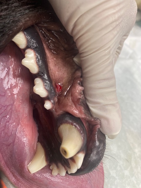
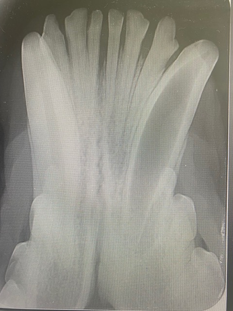
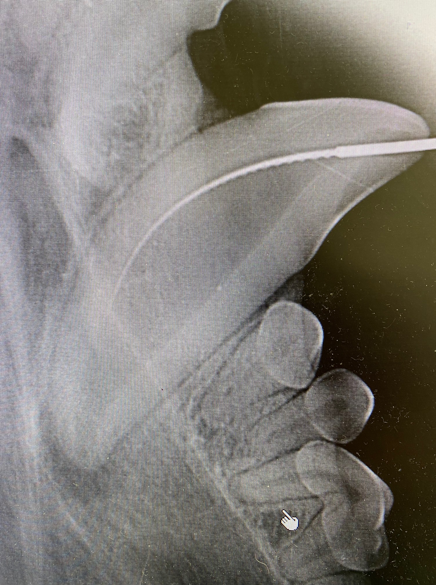
When under the anaesthetic the mouth could be properly examined, and a draining fistula identified at the point of the apex of the canine tooth, directly below 306. Pre op x-rays showed the the apex of the root of 304 was not closed. This means that most likely a traumatic incident occurred at the age of 9-12 months, causing the tooth to die and the normal development of the tooth to stop. As you can see in the x-ray, 304 has a much larger pulp than 404, indicating 304 is dead.
A root canal CAN NOT be done on this tooth as there is no “Stopper” at the apex of the pulp to contain the substances used to fill the tooth. (Note, however, that if the problem had been identified at the time, the tooth could have been filled with CaOH which can stimulate odontogenesis and the apex to close, meaning a root canal could be performed at a later date).
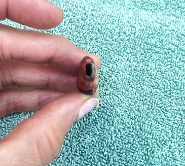
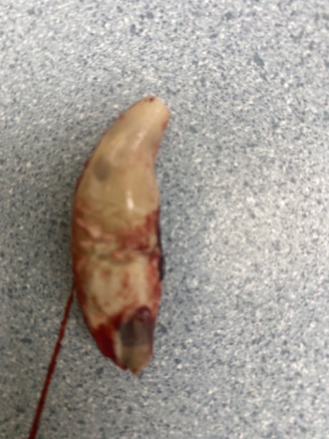
In this case, 304 had to be carefully extracted. This was done by making a gingival flap, burring away buccal and some lingual alveolar bone, and by using a vet tome, luxators, elevators and forceps. The photos show the open apex and thin dentine of 304 post extraction. A post op x-ray showed complete extraction of the root. The socket was curetted and flushed with copious amounts of saline. A periosteal releasing incision was made on the underside of the gingival flap and it was closed under no tension using 4/0 mono q absorbable suture material.
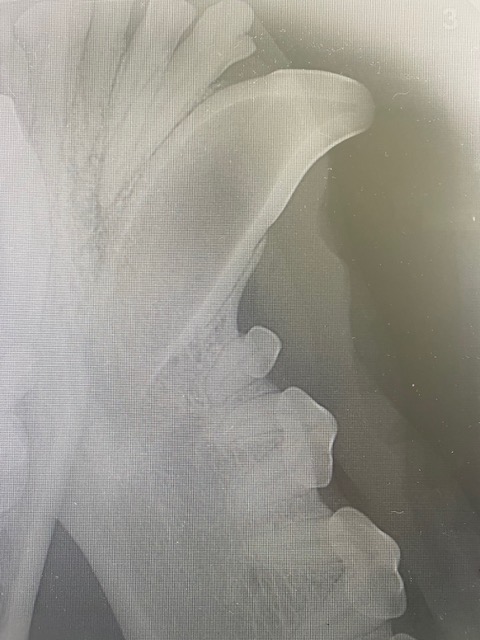
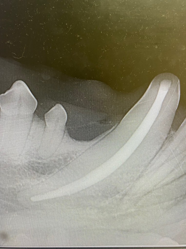
The above x-ray is an example of a young dog with a root apex that HAS closed. The dentine and periodontal ligament can be seen all the way around the apex of the tooth. This dog is likely just over 12-15 months old. A root canal CAN be done on this tooth as there is a “stopper” or end to the pulp cavity that will contain the substance used to fill the canal. There is also periapical lysis indicating infection is present.
The above tooth has had a root canal procedure performed. You can see the radiopaque filler (in this case flowable gutta percha with a GP point) fills the whole canal, and can be compressed down to where the pulp chamber ends.
