
This case was referred to me because the permanent left mandibular canine tooth (304) had not erupted into the mouth. X-ray from the referring vet revealed it was retained. History from a previous vet clinic revealed the deciduous mandibular canine teeth had been lingually displaced so had been extracted, and that the deciduous canine tooth on the left side (704) had fractured and the root left insitu.
It is most likely that during extraction of the deciduous tooth the crown was damaged and for this reason the permanent canine tooth on this side did not erupt.
Interestingly the permanent right canine tooth (404) was lingually displaced and a crown reduction and vital pulpotomy was performed on this tooth after the extraction of 304.
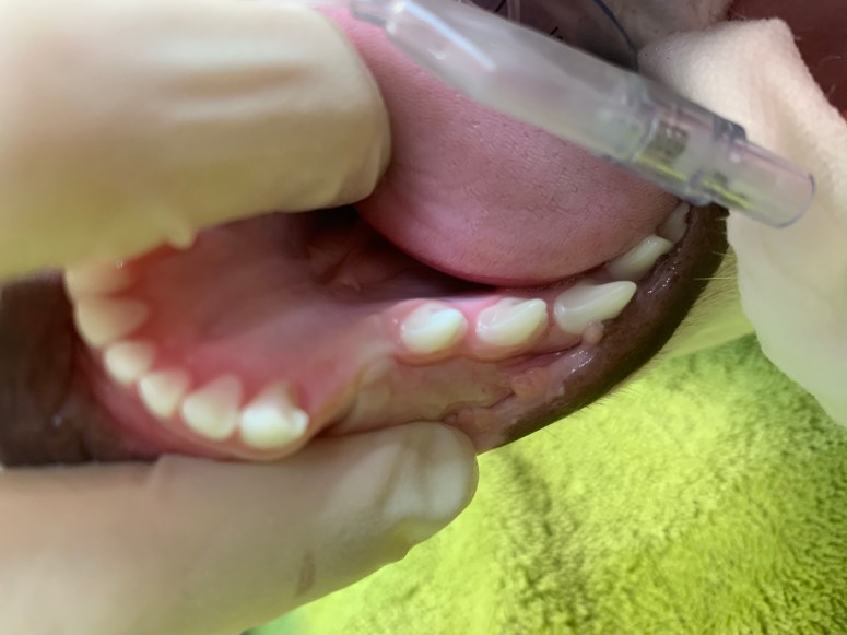
Pre op photo showing the area where the canine tooth should be 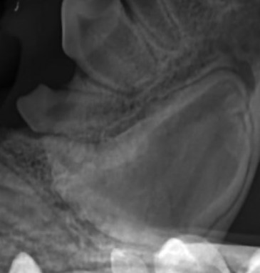
Abnormal crown structure of retained canine 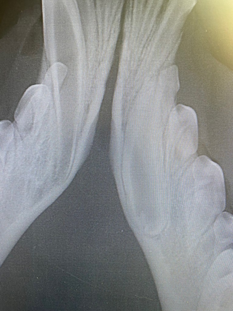
The retained canine tooth can be seen on x-ray very close to the mandibular cortex 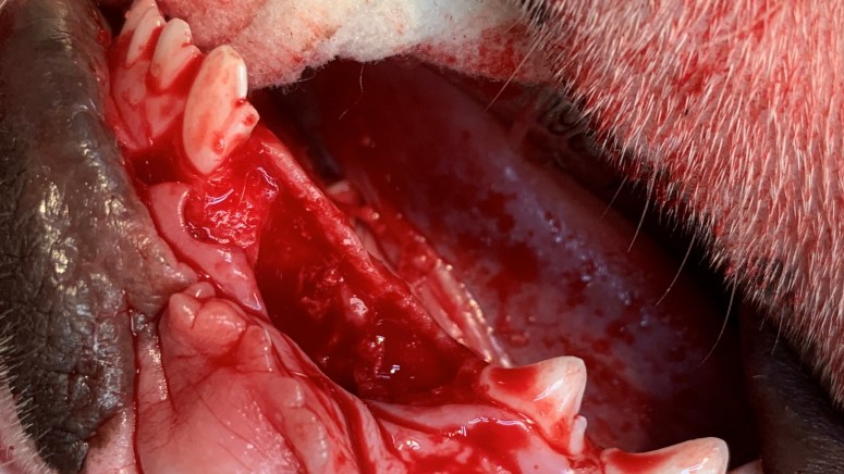
305 and 306 were extracted and bone burred away to allow removal of the retained canine 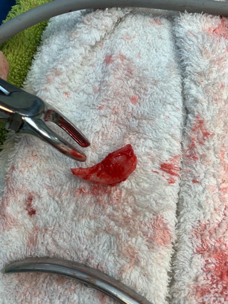
The retained canine tooth with very wide root and abnormal crown 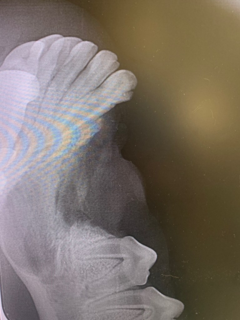
Post op x-ray after extraction
To extract the retained canine tooth alveolar bone was burred away and the two mesial premolars 305 and 306 extracted to allow visualization and access to 304. A vet tome instrument, luxators and elevators were then carefully used to stretch and lance the periodontal ligament around the root. Post extraction the socket was debrided and flushed with saline then a bone graft material (Synergy) was placed in the empty socket. The site was sutured using 4/0 absorbable suture material.
The dog was not allowed to chew on hard substances for 4 weeks to allow time for new mandibular bone to form and the jaw to strengthen. This is because the retained tooth was large and also sitting very close to the mandibular cortex.
A possible complication in this case was jaw fracture, so a gentle technique, time and patience were necessary during surgery. The vet tome was also instrumental in the successful result!
