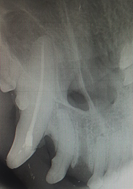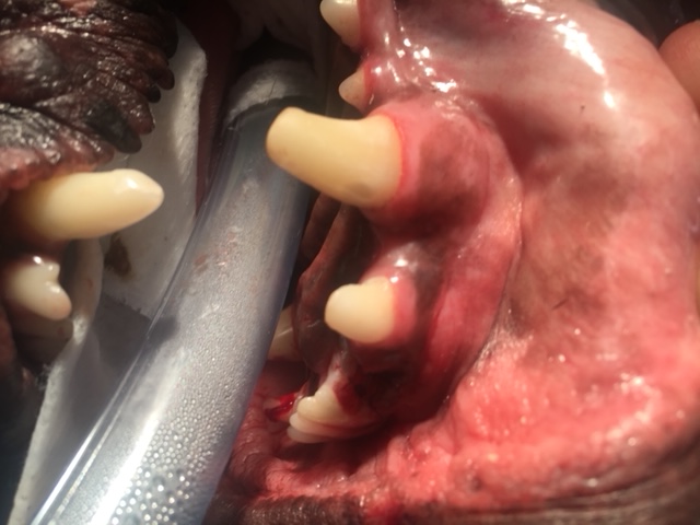

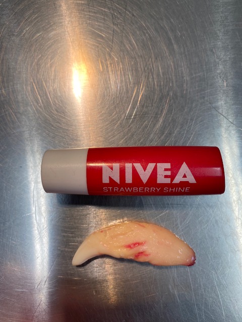
As you can see in the photos the canine tooth is massive. The crown, which is the part that extends into the mouth, is only a small part of this tooth. The root is large and held in the jaw by very a strong periodontal ligament that has fibres running from the tooth to the surrounding bone.
Structurally canine teeth are important as they hold the shape of the mouth, provide structure to the jaw, keep the tongue in place in the mouth (mandibular/lower canines) and are used in eating, holding and playing. If a canine tooth is fractured and the pulp is exposed, then the two options for a pet over 1 year old are a root canal or extraction. Under 1 year old the apex of the tooth may not be closed so a root canal may not be able to be done.
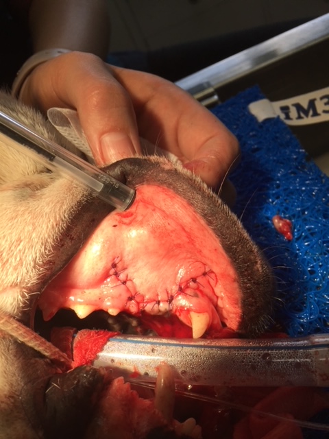
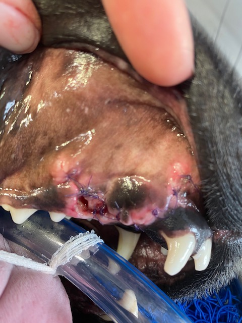
For an extraction, a large gingival flap is made and alevolar bone is burred away. The amount of bone burred away will be less if a vet-tome instrument is used. The tooth is then elevated and luxated out of the socket. When teeth are fractured (as opposed to periodontally diseased), the tooth is normally structurally sound so extraction of the tooth does require a fair amount of force. It is relatively traumatic. Post extraction the socket is flushed with saline and the gingival flap sutured closed. It is important the dog eats soft food and does not chew on anything hard while the gum is healing as wound breakdown can occur and the dog animal may need another anesthetic for resuturing the gingival flap.
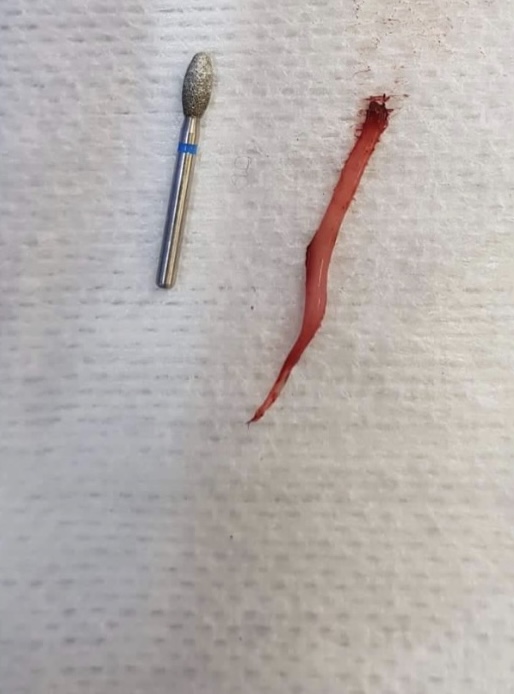
When a root canal operation is performed, a small access hole is made on the mesial surface of the tooth and the pulp is removed using files and solutions (bleach, EDTA, saline). Once cleaned and dried the pulp is filled with Gutta Percha (a sterile rubbery type substance that sets hard) and the access hold and fracture sites have a restoration placed on them. A follow up X-ray should be done 12 months after surgery to ensure no complications have occurred.
If possible I recommend keeping the canine teeth in the mouth especially in a young animal.