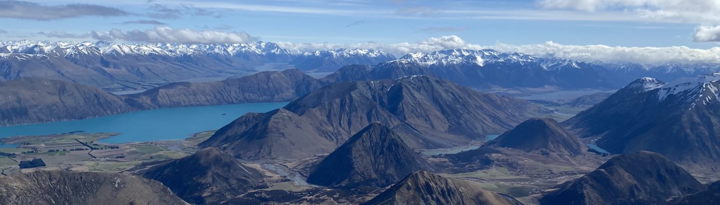

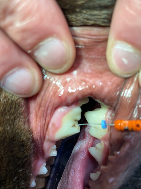
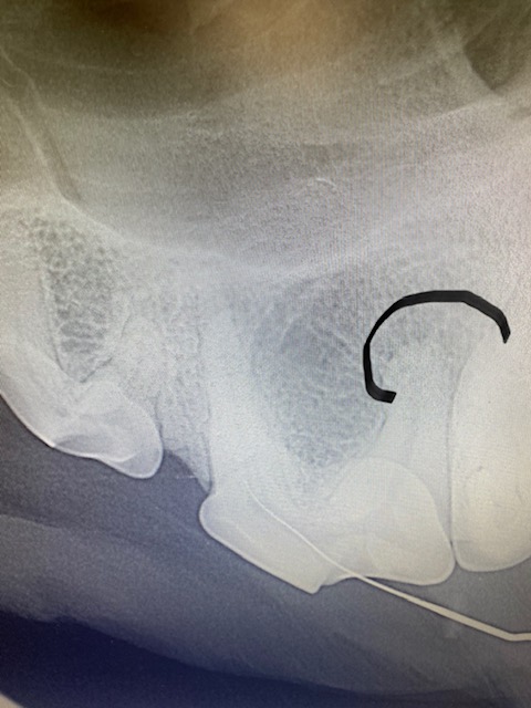
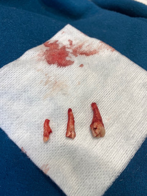
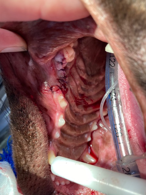
Tooth 208 (upper left premolar) had a complicated crown fracture. When under anesthetic a small pathfinder file was inserted into the pulp. A pre op X-ray showed a slightly lytic circular region around the distal root (black line outlies this). This tooth could have had a root canal operation performed if the owner had wanted to save the tooth, however in this instance an extraction procedure was elected. A maxillary nerve block using bupivicaine was performed, then the tooth was sectioned into 3 and each root removed separately. On the distal root a granuloma can be seen around the apex (this is what was visible on the X-ray). After extraction an X-ray was taken to confirm no root remnants remained and the alveolar bone was smoothed with an oval diamond head bur. The gingival flap sutured closed with simple interrupted 4/0 absorbable suture material with no tension. NSAID pain relief was given post. Antibiotics were not dispensed.